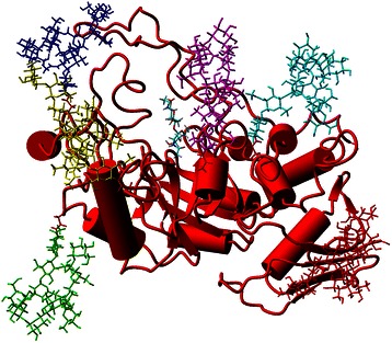
Figure 5.

Glycosylated model of TfHex. Side view of glycosylated, monomeric TfHex with carbohydrate antennae shown as stick models (red is connected to Asn 170, green – Asn 343, blue – Asn 378, yellow – Asn 433, magenta – Asn 453, cyan – Asn 527). Position of the natural substrate chitobiose is shown in the active site in stick representation colored by element colors.
