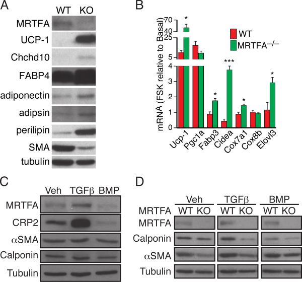
Figure 5. Cells from the stromal vascular fraction (SVF) of MRTFA–/– ING WAT express less smooth muscle-like features and undergo brown adipogenesis more extensively than WT SVF cells.
(A) Western blot analysis on BAT and WAT enriched proteins of adipocytes arising from hormonal induction of SVCs isolated from WT and MRTFA–/– inguinal depots.
(B) WT and MRTFA–/– SVC adipocytes were treated with or without forskolin (FSK) for 4 h prior to isolation of total RNA for analysis. Values are presented as fold change in response to treatment with FSK in WT and MRTFA–/– adipocytes.
(C and D) Subconfluent SVCs from ING and EPI WAT of WT and/or MRTFA–/– mice were exposed to TGFβ1 or BMP7 for 2 d before reaching confluence and subjected to Western blot analysis.

