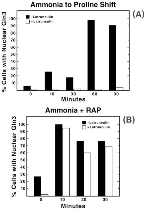
Fig. 3. Quantitation of Gln3 partitioning between the nucleus and cytoplasm in cells transferred from minimal-ammonia to -proline medium (panel A) or after treatment of ammonia-grown cells with rapamycin (RAP; panel B).
The experimental format used to transfer cells from glucose-ammonia to -proline medium was as described in Fig. 2. For rapamycin treatment, cultures (TB123) were grown to mid-log phase in 2% glucose, YNB (without amino acids or ammonium sulfate), and 0.1% ammonia sulfate and pretreated with 150 μM latrunculin B or carrier (ethanol) as described in Fig. 2. Aliquots were then removed just before the addition of 200 ng/ml rapamycin (0 min) and at the indicated times after the addition. Samples were processed for indirect immunofluorescence as in Fig. 2.

