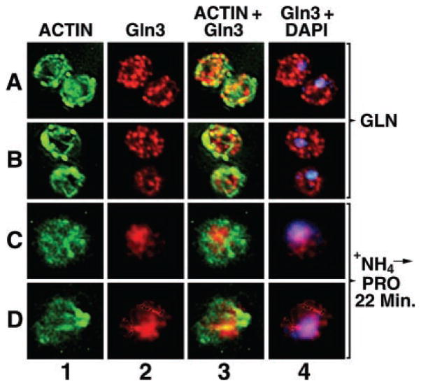
Fig. 9. Intracellular localization of Gln3 and actin.
Cultures (TB123) were grown to mid-log in 2% glucose, YNB-glutamine medium (rows A and B) or were transferred from glucose-ammonia to -proline medium, incubated at 30 °C for 22 min (rows C and D), and processed for indirect immunofluorescence. The localization of actin, Gln3-Myc13, and nuclei was determined by staining with phalloidin (columns 1 and 3, green), monoclonal antibody 9E10 anti-Myc (columns 2–4, red), and DAPI (column 4, blue), respectively. Merged images (column 3) show the overlap (column 3, yellow) of Gln3 and actin or Gln3 and DAPI-positive material (column 4, pink). Micrographs were imaged using a Zeiss Axioplan 2 imaging microscope. 0.4-μm sections were collected as a Z-stack, and one image from the center of that stack is shown. Images were deconvolved with AxioVision 3.0 software using the constrained iterative algorithm.

