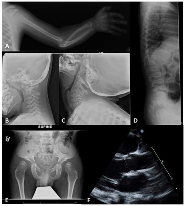
Figure 2.
A. Left shoulder and elbow: Proximal humeral flattening, subluxation of the humeral head, dysplastic glenoid, dysplastic distal humerus with dislocated elbow, dysplastic radial head, bowing of radius and ulna, and functionally fused radio-ulnar joint.
B., C. C-spine upright flexion (B) and extension (C): Atlantoaxial and atlantooccipital instability.
D. Spine Xray lateral: Gibbus deformity at the L1 level, and gracile ribs.
E. Hip: Flat acetabular angles, broad and flattened iliac wings, and a lacy trabecular pattern.
F. Echocardiogram: Mild dilation of the aortic root and main pulmonary artery.

