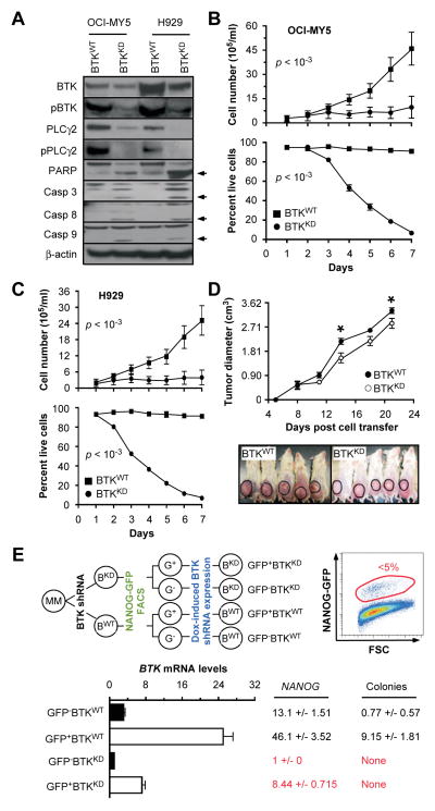
Figure 5. Down regulation of BTK in myeloma cells mitigates growth, survival, and stemness.
(A) Western blots of OCI-MY5 and H929 myeloma cells that either under-express BTK due to lentiviral transduction of a BTK–targeted shRNA “knock down” construct (BTKKD) or express BTK at normal levels due to transduction of a non-targeted or “scrambled” shRNA (BTKWT). PARP indicates poly (ADP-ribose) polymerase. Casp3, 8 and 9 denote three different members of the apoptosis-related cysteine peptidase family of caspase proteins.
(B) Line graphs presenting changes in cell number (top) and cell viability (bottom) of BTKKD and BTKWT OCI-MY5 cells grown for 7 days in vitro.
(C) Same as panel B except H929 cells were used.
(D) Time course of tumor growth in NOD-SCID mice, showing that BTKKD cells expand less vigorously in vivo than BTKWT cells.
(E) Evidence indicating that BTK and NANOG are co-regulated in myeloma, and that the BTK-NANOG axis promotes clonogenicity of myeloma cells.

