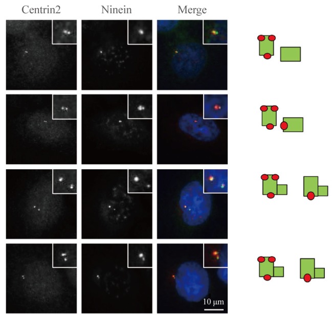
Fig. 3. Ninein distribution within the centriole. Asynchronous HeLa cells were coimmunostained with centrin-2 (green) and ninein (red) antibodies. DNA was stained with 4',6-diamidino-2-phenylindole. Scale bar=10 µm. Insets are magnified views of the centrosomes. The staining patterns of ninein on centrioles were taken from the right side.

