. 2015 Mar 27;30(1):53–57. doi: 10.3803/EnM.2015.30.1.53

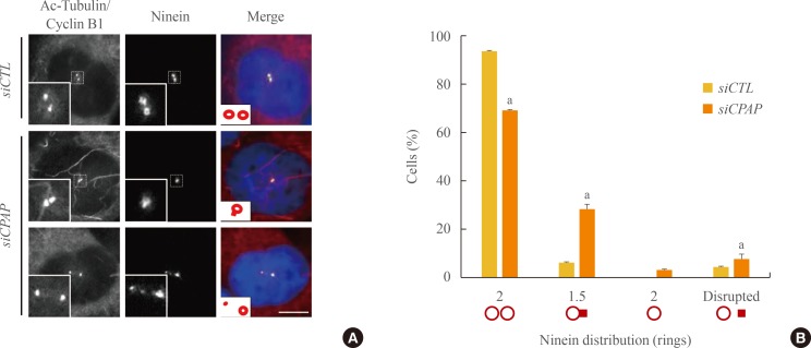
Fig. 4. Centrosomal ninein distribution in centrosomal P4.1-associated protein (CPAP)-depleted mitotic cells. (A) The cell cycle of CPAP-depleted cells was synchronized during the G2 phase using a double thymidine block and release. The cells were placed on ice for 90 minutes to disrupt microtubules, and coimmunostained with acetylated-tubulin (red), cyclin B1 (red), and ninein (green) antibodies. DNA was stained with 4',6-diamidino-2-phenylindole. Scale bar=10 µm. (B) Ninein staining patterns at the spindle poles were categorized into four groups: paired ring; ring with a bar; single ring; and disrupted. More than 300 cells were analyzed across three independent experiments. The data are means±SE. aP<0.05 was considered statistically significant compared to the controls.

Fig. 4