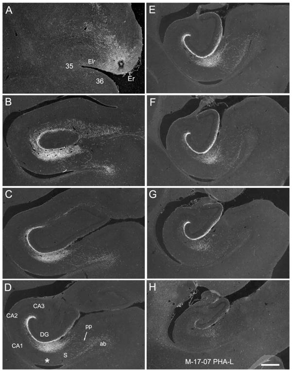
Figure 11.
A–H: Darkfield photomicrographs illustrating the injection (A) and anterogradely transported label in the hippocampal formation in case M-17-07. A: The PHA-L injection site is rostrally placed and involves primarily layers III and II of the rostral part of the entorhinal cortex (Er). Anterogradely transported fibers travel in the angular bundle (ab in D) traverse the subiculum (S) in the perforant path (pp in D), and innervate the dentate gyrus, CA1, and subiculum. Labeling is very extensive rostrocaudally and extends from near the rostral pole of the dentate gyrus (B) to nearly its caudal end (H). The injection is very rostrally placed, and fibers terminate most heavily in the outer third of the molecular layer of the dentate gyrus and at the border between CA1 and the subiculum (asterisk in D). Scale bar = 1 mm in H (applies to A–H).

