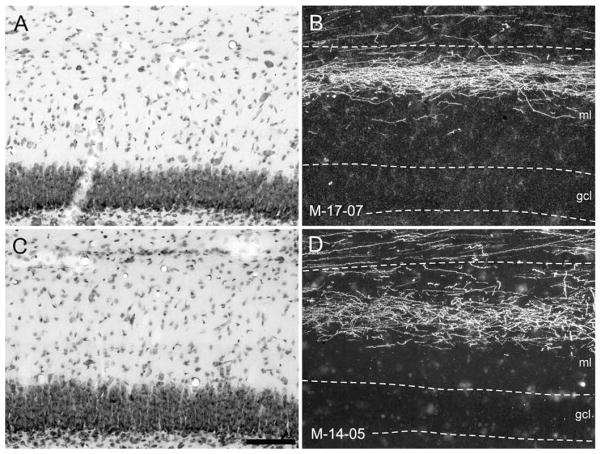
Figure 13.
A–D: Higher magnification photomicrographs of coronal sections showing the laminar distribution of anterograde labeling in the molecular layer (ml) of the dentate gyrus following the tracer injections in the rostral (B, PHA-L in case M-17-07) and caudal (D, BDA in case M-14-05) parts of the entorhinal cortex in infant monkeys. These are the same cases illustrated at lower magnification in Figure 12. Adjacent Nissl-stained sections for B and D are shown in A and C, respectively. The superficial and deep borders of the molecular layer of the dentate gyrus and the granule cell layer (gcl) are indicated by dashed lines. Note that labeled axons and terminals are preferentially located in the superficial portion of the molecular layer of the dentate gyrus following the rostral injection (B), whereas labeled axons and terminals are preferentially located in the mid-portion of the molecular layer following the caudal injection. Scale bar = 100 μm in C (applies to A–D).

