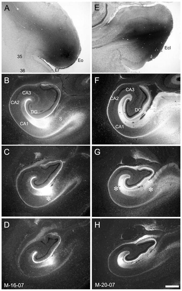
Figure 7.
A,E: Brightfield photomicrographs of the 3H-amino acid injections in cases M-16-07 (A) and M-20-07 (E). The injection in M-16-07 is near the rostral pole of the entorhinal cortex and involves fields Eo and Er. The injection in case M-20-07 is near the caudal pole of the entorhinal cortex and involves the caudal limiting field (Ecl). B–D,F–H: Darkfield photomicrographs of coronal sections of the infant monkey hippocampal formation arranged from rostral (B and F) to caudal (D and H) showing the distribution of anterogradely labeled projections in cases M-16-07 and M-20-07, respectively. In both cases, labeling is distributed extensively through the rostrocaudal extent of the dentate gyrus. However, in M-16-07, the heaviest labeling is in the outer third of the molecular layer of the dentate gyrus, whereas in case M-20-07 labeling is much heavier in the middle third of the molecular layer. Differences are also seen in the topography of the projection to CA1 and the subiculum as indicated by the asterisks in C and G. The rostral injection in M-16-07 leads to labeling at the border of these two fields, whereas the caudal injection in M-20-07 leads to heaviest labeling in CA1 close to CA2 and in the subiculum close to the presubiculum. For abbreviations, see Figure 1 legend. Additional abbreviations: 35 and 36, areas 35 and 36 of the perirhinal cortex. Scale bar = 1 mm in H (applies to A–H).

