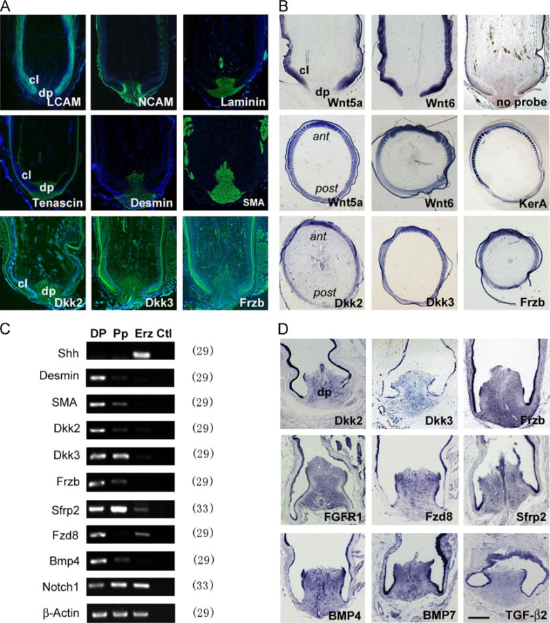
Fig. 2.

Gene expression analysis in the feather follicle. (A) Marker gene expression shown by immunofluorescence (green). LCAM marks the feather epithelium. NCAM marks the DP/dermal sheath and weakly the feather branching epithelium. Laminin marks the DP and vessel walls. Tenascin marks the DP/dermal sheath. Desmin is more DP specific, and SMA marks the DP and vessel walls. Dkk2/Dkk3/Frzb is enriched in the DP, presents in the pulp but less in the epithelium. Some unspecific staining is found in the keratinized feather sheath. (B) Expression of Wnt ligands and inhibitors in the feather follicles shown by in situ hybridization. Notice Wnt5a and Wnt6 appear primarily in the epithelium. A gradient distribution pattern is detected for Wnt5a/Wnt6, Dkk2/Dkk3/Frzb and feather keratin A in the Erz region. A control staining with no probe is also shown. (C) Semi-quantitative RT-PCR and (D) in situ hybridization analysis of gene expression in the feather follicles. No template reactions are used as control for PCR analysis, and equal amount of RNAs are monitored by β-Actin gene expression. The number after each gene indicates PCR cycles. dp, dermal papilla; cl, collar; ant, anterior where the rachis locates; post, posterior as opposite to the rachis position. Bar = 1 mm in A and B, and 0.5 mm in D (shown in D).
