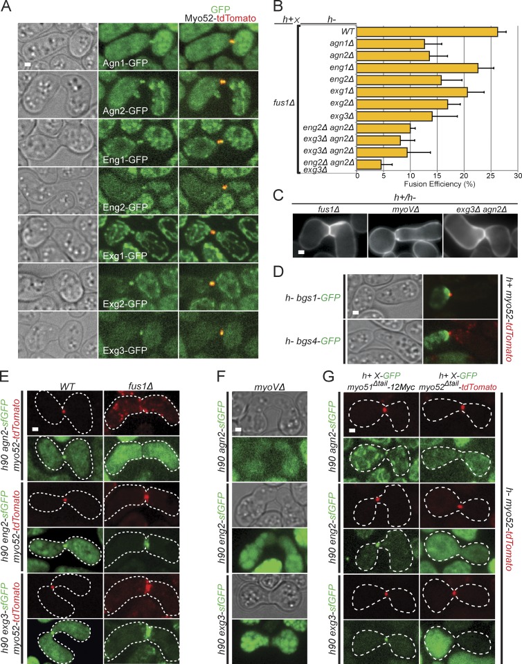
Figure 5.
Cell wall degradation enzymes focalize at the fusion focus. (A) Crosses of heterothallic h− and h+ myo52-tdTomato strains expressing GFP-tagged glucanases as indicated. All glucanases localize to the fusion focus. (B) Fusion efficiency of crosses between h+ fus1Δ strains and h− single, double, and triple glucanase deletion strains. n > 100. (C) Calcofluor images of nonfusing pairs in heterothallic crosses of h+ fus1Δ, myoVΔ, and agn2Δ eng2Δ to h− fus1Δ, myoVΔ, and agn2Δ eng2Δ, as indicated. This shows presence of cell wall at the cell–cell contact. (D) Time-averaged projections over 10 s of crosses of heterothallic h− bgs1-GFP (top) and bgs4-GFP (bottom) to h+ myo52-tdTomato, imaged every second. Cell wall synthases localize as a crescent at the fusion site. (E) Homothallic h90 wild type (left) and fus1Δ (right) myo52-tdTomato strains coexpressing sfGFP-tagged versions of Agn2, Eng2, and Exg3. Cell wall glucanases colocalize at fusion site with Myo52 either as a dot in wild type or as a crescent in fusion-deficient fus1Δ cells. Cell outlines are shown with dotted lines. (F) Homothallic h90 myo51Δ myo52Δ (myoVΔ) strains coexpressing sfGFP-tagged versions of Agn2, Eng2, and Exg3. Glucanases are not detected at fusion site. (G) Heterothallic h+ myo51Δtail-12Myc (left) and h+ myo52Δtail-tdTomato (right) strains coexpressing sfGFP-tagged versions of Agn2, Eng2, and Exg3 crossed to h− myo52-tdTomato cells. Cell wall glucanases localize at the fusion site in myo51Δtail mutants but not, or are highly reduced, in myo52Δtail. WT, wild type. Error bars are standard deviations. Cell outlines are shown with dotted lines. Bars, 1 µm.

