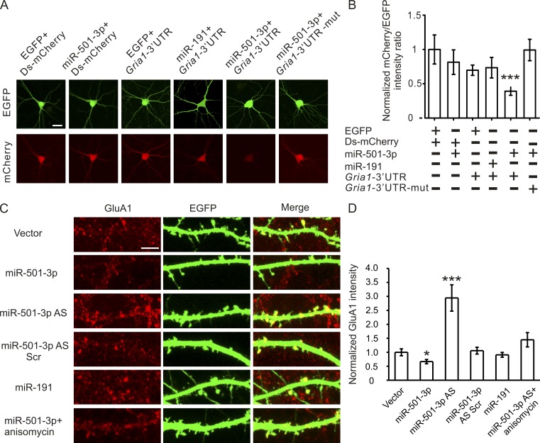
Figure 2.
Gria1 is a physiological target of miR-501-3p. Cultured hippocampal neurons were transfected with designated constructs at 14 DIV and imaged at 17 DIV. (A) Representative images of neurons cotransfected with the miRNA and the reporter construct. (B) Quantification of A; n = 12–15 neurons for each group. (C) Representative images of dendrites from transfected neurons stained with the GluA1 antibody. (D) Quantification of C; n = 14–20 neurons for each condition. Data are presented as mean ± SEM; one-way ANOVA was used for statistical analysis among different groups; P < 0.05. Mann-Whitney U test is used for statistical analysis; *, P < 0.05; ***, P < 0.005. Bars: (A) 20 µm; (C) 5 µm.

