Figure 3.
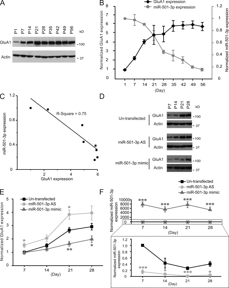
The expression of miR-501-3p and GluA1 is inversely correlated during development. Proteins and total RNAs were isolated from the hippocampus of rats and cultured hippocampal neurons at indicated ages for immunoblotting (A, B, D, and E) or qRT-PCR (B and F). (A and D) Representative blots. (B and E) Quantification of A and D; n = 3–8 rats or cultures for each age. (B and F) qRT-PCR analysis of miR-501-3p expression in the rat hippocampus (B) and cultured hippocampal neurons (F); n = 3–8 rats or cultures for each age group. (C) Correlation between expression levels of miR-501-3p and GluA1 at different ages in the rat hippocampus. Data are presented as mean ± SEM; one-way ANOVA was used for statistical analysis among different groups; Mann-Whitney U test was used for statistical analysis; *, P < 0.05; **, P < 0.01; ***, P < 0.005.

