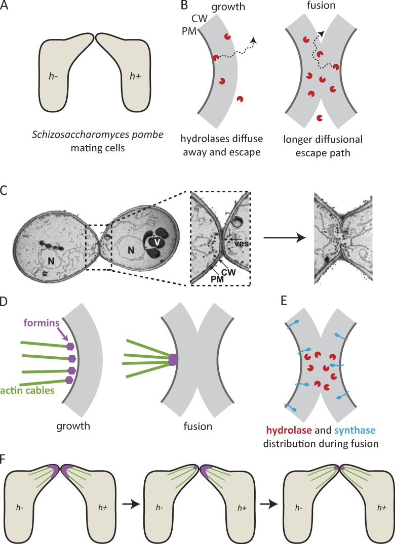
Figure 1.
Cell fusion during yeast mating: focus and communication. (A) Mating fission yeast cells grow projections toward each other and fuse at the point of contact. (B, left) Secreted hydrolases weaken the rigid cell wall to enable expansion, and rapidly diffuse away. (B, right) At a point of cell–cell contact, diffusional escape paths are longer, so hydrolases build up. (C) Focused delivery of secretory vesicles (ves) in mating budding yeast after contact. The image is adapted from Gammie et al. (1998), © The American Society for Cell Biology. (D) Actin cables during growth of the projection (left) and in the fusion focus (right). (E) Distribution of hydrolases and synthases in fusing cells. (F) The fusion focus forms first in the h− mating partner and then in the h+ mating partner. CW, cell wall; PM, plasma membrane; N, nucleus; V, vacuole.

