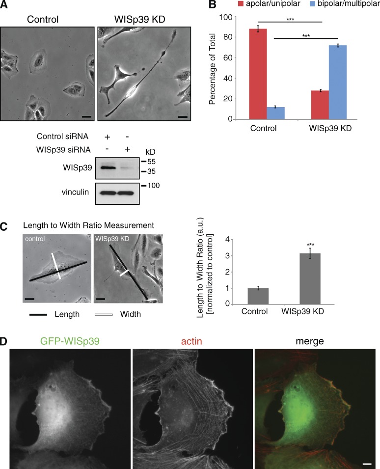
Figure 1.
Loss of WISp39 results in elongated cell morphology. (A) Subconfluent U2OS cells were transfected with either control (left) or human WISp39 siRNA (WISp39 KD; right) for 48 h and representative 20× phase images were taken. Western blot shows KD of WISp39 at 48 h. Vinculin is a loading control. (B) Depletion of WISp39 changes cell polarity. Cells were scored as either apolar (round)/unipolar (one lamellipodium) or bipolar/multipolar (two or more lamellipodia) and are presented as a percentage of total cells scored from more than five independent experiments. n = 1,165 control cells; n = 1,483 WISp39 KD cells. Data represent the means ± SD. Student’s t test; ***, P ≤ 0.001. (C) Quantitation of the maximum length to maximum width ratio of control cells versus WISp39 KD cells from an experiment performed as in A. Number of cells scored per group is 50 from more than three independent experiments. Data represent the means ± SD. Student’s t test; ***, P ≤ 0.001. a.u., arbitrary unit. (D) Cytoplasmic WISp39 localizes to actin filaments at the leading edge. U2OS cells were transfected with GFP-WISp39, fixed with paraformaldehyde, and stained with rhodamine-phalloidin (actin). Bars, 10 µm.

