. 2015 Jan 29;4:e05378. doi: 10.7554/eLife.05378

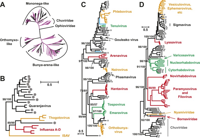
Figure 3. Evolutionary history of negative-sense RNA viruses based on RdRp.

This is initially displayed in an unrooted maximum likelihood (ML) tree including all major groups of negative-sense RNA viruses (A). Separate and more detailed ML phylogenies are then shown for the Orthomyxoviridae-like (B), Bunya-Arenaviridae-like (C), and Mononegavirales-like viruses (D). In all the phylogenies, the RdRp sequences described here from arthropods are either shaded purple or marked with solid gray circles. The names of previously defined genera/families are labeled to the right of the phylogenies. Based on their host types, the branches are shaded red (vertebrate-specific), yellow (vertebrate and arthropod), green (plant and arthropod), blue (non-arthropod invertebrates), or black (arthropod only). For clarity, statistical supports (i.e., approximate likelihood-ratio test (aLRT) with Shimodaira–Hasegawa-like procedure/posterior probabilities) are shown for key internal nodes only.

DOI: http://dx.doi.org/10.7554/eLife.05378.009

Figure 3.

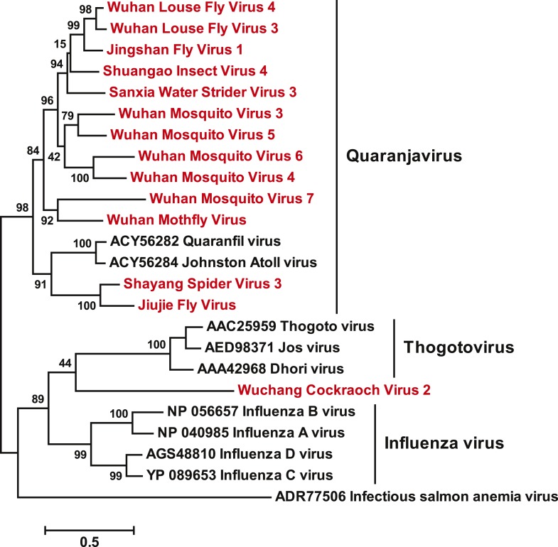
Figure 3—figure supplement 1. A fully labeled ML phylogeny for Orthomyxoviridae-like viruses.

Figure 3—figure supplement 1.

The phylogeny is reconstructed using RdRp alignments. Statistical support from the approximate likelihood-ratio test (aLRT) is shown on each node of the tree. The names of the viruses discovered in this study are shown in red. The names of reference sequences, which contain both the GenBank accession number and the virus species name, are shown in black. The names of previously defined genera/families are shown to the right of the phylogenies.
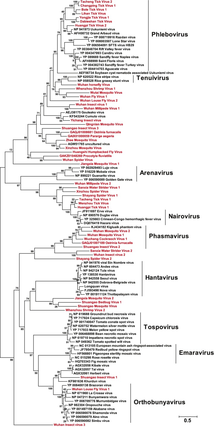
Figure 3—figure supplement 2. A fully labeled ML phylogeny for Bunya-Arenaviridae-like viruses.

Figure 3—figure supplement 2.

The phylogeny is reconstructed using RdRp alignments. Statistical support from the aLRT is shown on each node of the tree. The names of the viruses discovered in this study are shown in red. The names of reference sequences, which contain both the GenBank accession number and the virus species name, are shown in black. The names of previously defined genera/families are shown to the right of the phylogenies.
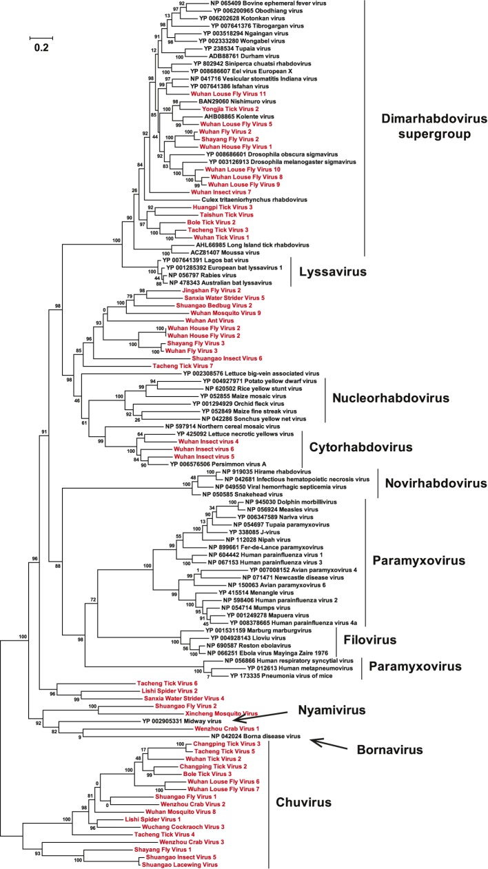
Figure 3—figure supplement 3. A fully labeled ML phylogeny for Mononegavirales-like viruses.

Figure 3—figure supplement 3.

The phylogeny is reconstructed using RdRp alignments. Statistical support from the aLRT is shown on each node of the tree. The names of the viruses discovered in this study are shown in red. The names of reference sequences, which contain both the GenBank accession number and the virus species name, are shown in black. The names of previously defined genera/families are shown to the right of the phylogenies.