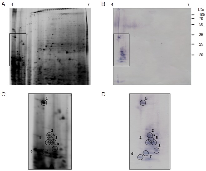
Fig. 5.

Representative 2-DE images of the whole body proteins from partially fed female ticks of H. longicornis. (A) Silver stained gel. (B) Immunoblot probed with polyclonal antibodies against H. longicornis. Proteins (240 μg) were focused on pH 4-7 IPG strips (13 cm) and were then separated by SDS/12.5% PAGE. Reference molecular masses are indicated on the right of the gel. 2-DE immunoblot shows the antigenic spots revealed by a pool of sera from rabbits experimentally infested with H. longicornis. The proteins of interest are marked in the image (rectangles). (C, D) Magnifications of panels A and B. In D, antigenic spots are indicated by black numbered circles and are equivalent to the proteins identified in panel.
