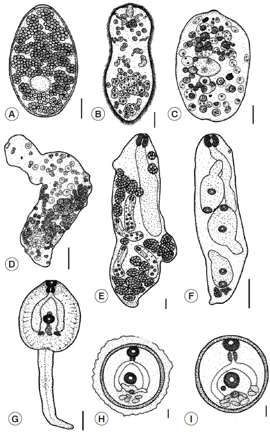
Fig. 4.

Illustration demonstrating the larval morphology of F. gigantica. (A) Egg. (B) Miracidium. (C) Young sporocyst. (D) Mature sporocyst. (E) Mother redia. (F) Daughter redia. (G) Cercaria. (H) Encapsulated metacercaria. (I) Metacercaria. Scale bars (A-D)=0.03 mm; (E-G)=0.1 mm; (H-I)=0.05 mm.
