Figure 1.

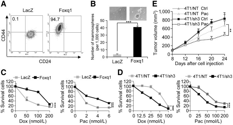
Foxq1-induced EMT, stemness traits, and chemoresistance in mammary epithelial cells and breast cancer cells. A, FACS analysis of cell-surface markers, CD44 and CD24, in HMLE cells with Foxq1 gene and LacZ control overexpression. B, in vitro quantification of mammospheres formed by cells described in A. The data are reported as the number of mammospheres formed/1,000 seeded cells ± SEM, compared with control (two experiments performed in triplicate; ***, P < 0,001). C and D, chemoresistance of HMLE cells (C; with or without Foxq1 overexpression) and 4T1 cells (D; with or without Foxq1 knockdown) was analyzed by an MTT assay after treatment with doxorubicin or paclitaxel as indicated for 24 hours. Percentages of live cells compared with the nontreatment control are shown (*, P < 0.05; **, P < 0.01). E, knockdown of Foxq1 led to significant sensitization of 4T1 cells to paclitaxel treatment in vivo (*, P < 0.05; **, P < 0.01).
