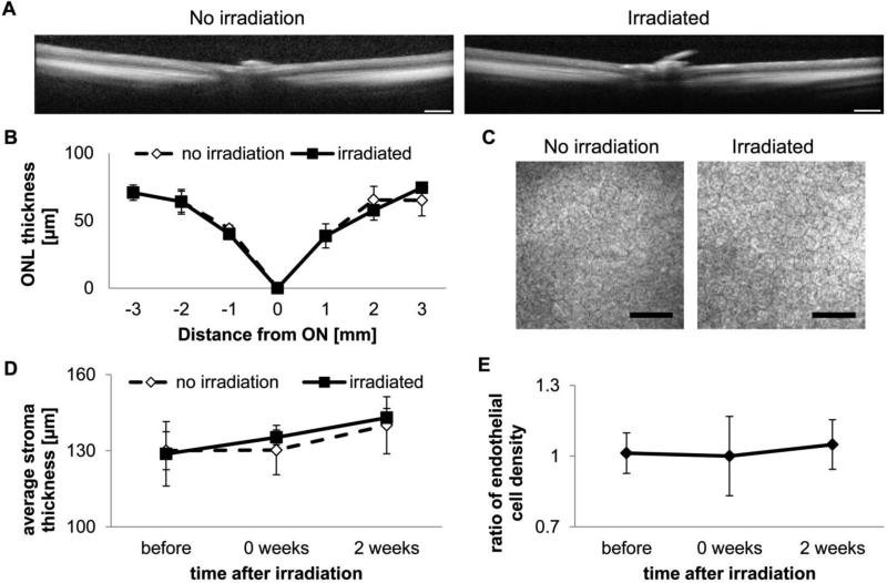
Figure 3. Irradiation required for release does not damage the eye.
A, Representative optical coherence tomography (OCT) scans through the retina (scale bar = 0.5 mm). B, Quantification of outer nuclear layer (ONL) thickness (ON – optical nerve, positive – towards temporal quadrant, negative – towards nasal quadrant; n = 3). C, Representative confocal tomographs of the corneal endothelium 2 weeks post-irradiation (scale bar = 50 μm). D, Quantification of stromal thickness from volume cornea tomographs (n = 4). E, Quantification of endothelial cell count ratio (n = 4).

