
Figure 3.
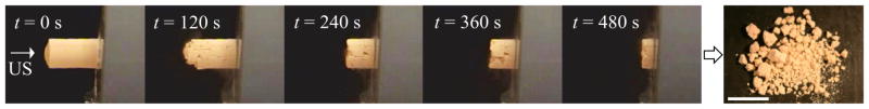
Photographic sequence of an artificial stone during exposure to 170 kHz bursts with pf = 6.5 MPa over 8 minutes. Ultrasound (US) burst waves are incident on the stone from the left. The photograph to the right shows the fragments generated after 8 minutes of exposure. The scale bar is 1 cm.