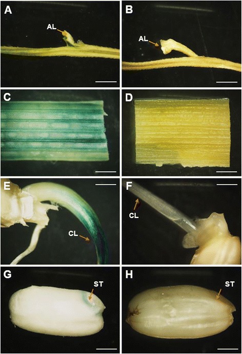
Figure 4.

GUS histochemical staining results in different tissues driven by pSH4. GUS-staining (blue dye) of different tissues in pSH4-GUS transgenic plants (A, C, E, and G) and non-transgenic controls (B, D, F, and H). A and B: branches of panicle, showing the seed-pedicel junction region (AL, indicated by arrows); C and D: stems (internodes); E and F: coleoptiles (CL, indicated by arrows) from germinated seeds; G and H: Dehusked mature seeds, showing scutella (ST, indicated by arrows). Bar = 1mm.
