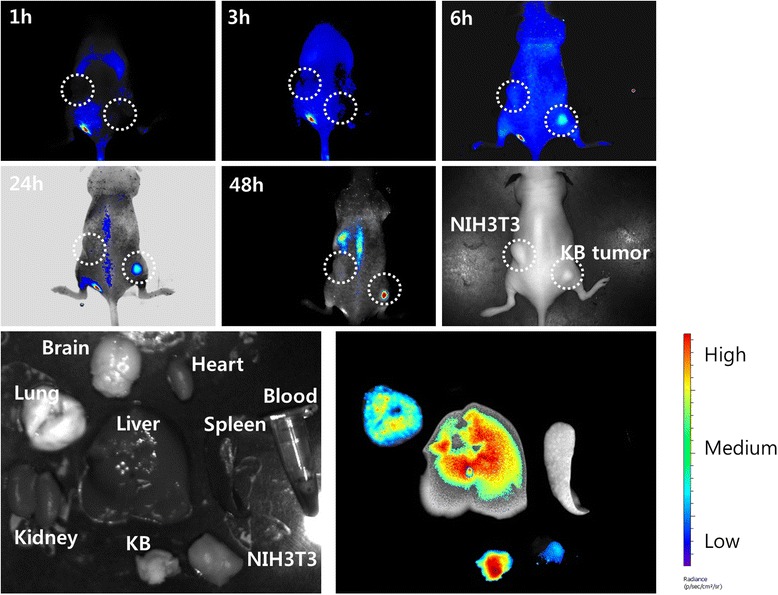
Figure 7.

The NIR fluorescence imaging study. BALb/C nude mouse bearing an NIH3T3 (left side) and KB tumor (right side) was used. NIR-dye-conjugated nanoparticles (20 mg/kg) were intravenously injected via the tail vein of the mouse. The major organs of the mice were taken 48 h after administration. The data shown as the mean ± SD (n = 4).
