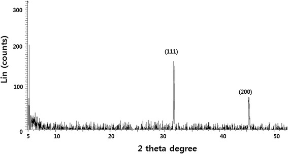
Figure 2.

XRD pattern of AgNPs. A representative X-ray diffraction (XRD) pattern of silver nanoparticles formed after reaction of leaf extract with 1 mM of silver nitrate (AgNO3) for 60 min at 60°C. The XRD pattern shows two intense peaks in the whole spectrum of 2θ values ranging from 20 to 70. The intense peaks were observed at 2θ values of 31.9 and 45.31 corresponding to (111) and (200) planes for silver, respectively.
