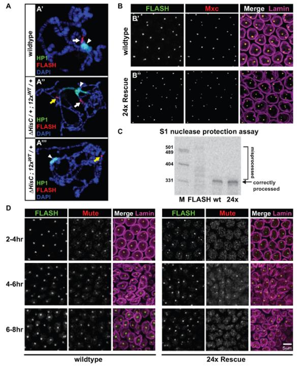
Figure 4. The transgenic histone gene locus assembles an HLB that accurately processes histone transcripts.
(A) Confocal images of salivary gland polytene chromosome squashes stained for FLASH (red), HP1 (green), and DAPI (blue) for the three indicated genotypes. White arrow: endogenous HLB; white arrowhead: chromocenter; yellow arrow: transgenic HLB. (B) Confocal images of blastoderm stage embryos stained for FLASH (green), Mxc (red), and Lamin (magenta) for the two indicated genotypes. (C) Phosphorimager scan of S1 nuclease protection assay performed on total RNA from three genotypes: FLASHPBac/FLASHDf; Oregon R (wt, 4-6hrs); 24× Rescue (24×, 4-6hrs). M: Markers. (D) Confocal images of embryos at 2-4hrs, 4-6hrs, and 6-8hrs stained for FLASH (green), Mute (red) and Lamin (magenta) for wild type and 24× Rescue embryos. For B and D, the maximum projection of four 0.5-micron slices is shown.

