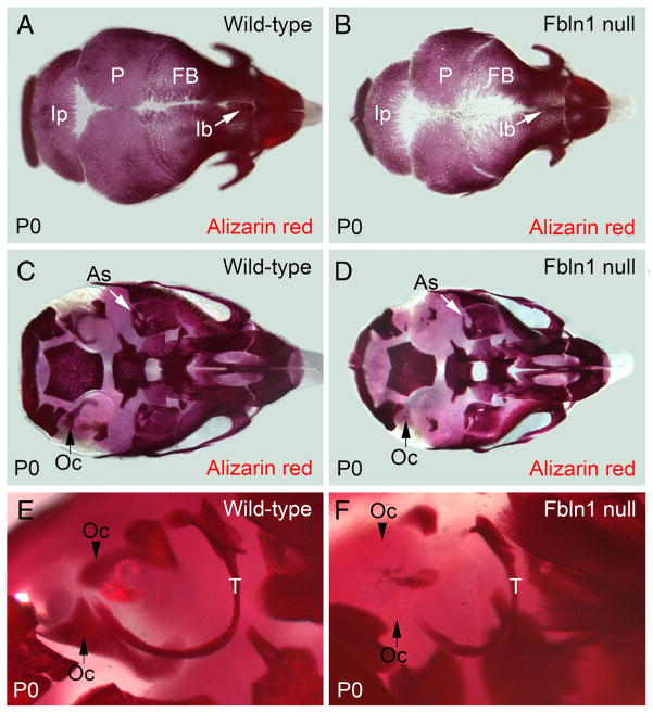
Fig. 1.
Fbln1 null neonates have reduced alizarin red staining of membranous bones and endochondral bones in the cranium. A and B show that alizarin red staining in the membranous frontal, parietal and interparietal bones from a P0 Fbln1 null neonate (B) is reduced as compared to P0 wild-type (A). Arrow in B points to the interfrontal bone in P0 Fbln1 null that is reduced compared to P0 wild-type. C and D show reduced alizarin red staining of the otic capsule from P0 Fbln1 null (D) as compared to P0 wild-type (C). Arrow (black) in C points to a bone in the otic capsule that is absent in the P0 Fbln1 null (black arrow in D). Arrow (white) shows alisphenoid bone in P0 Fbln1 null that has reduced alizarin red staining. E and F are higher magnification views of the otic region in P0 wild-type and P0 Fbln1 null skull showing reduced ossification of the otic capsule and tympanic ring bone (T) in P0 Fbln1 null. Arrowhead in E points to a bone in P0 wild-type otic capsule that is absent in the P0 Fbln1 null otic capsule. As, alisphenoid bone; FB, frontal bone; Ib, interfrontal bone; Ip, interparietal bone; Oc, otic capsule; P, parietal bone; T, tympanic ring bone.

