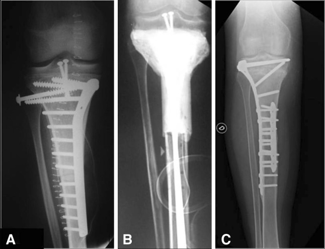
Fig. 4A–C.

A 17-year old male patient had an osteosarcoma of the right proximal tibia. He underwent reconstruction with an intercalary tibia allograft after transepiphyseal resection of a metadiaphyseal osteosarcoma. (A) An AP radiograph of the right proximal tibia obtained immediately after resection of the tumor and reconstruction with an allograft shows fixation of the allograft to the host bone with a medial buttress proximal plate and two screws in the remaining epiphysis. (B) An AP radiograph of the right proximal tibia 1 year after reconstruction shows a temporary antibiotic-impregnated cement spacer that replaced the original allograft owing to a deep infection. (C) Seven years postoperatively, the patient’s AP radiograph of the right tibia is shown. The patient underwent reconstruction with a second allograft with a new lateral long plate (proximal tibia locking compression plate) that covers both osteotomies and addition of a short medial plate in the distal osteotomy.
