
Figure 2.
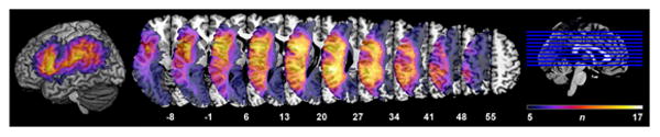
All patient lesions (n = 31) displayed on the Colin27 template in MNI standardized space. Color bar represents the number of patients with lesions at a particular voxel (min = 5; max = 17). Voxels in which fewer than 5 patients had a lesion were not included in VLSM analyses and so are not displayed.