
Figure 1.
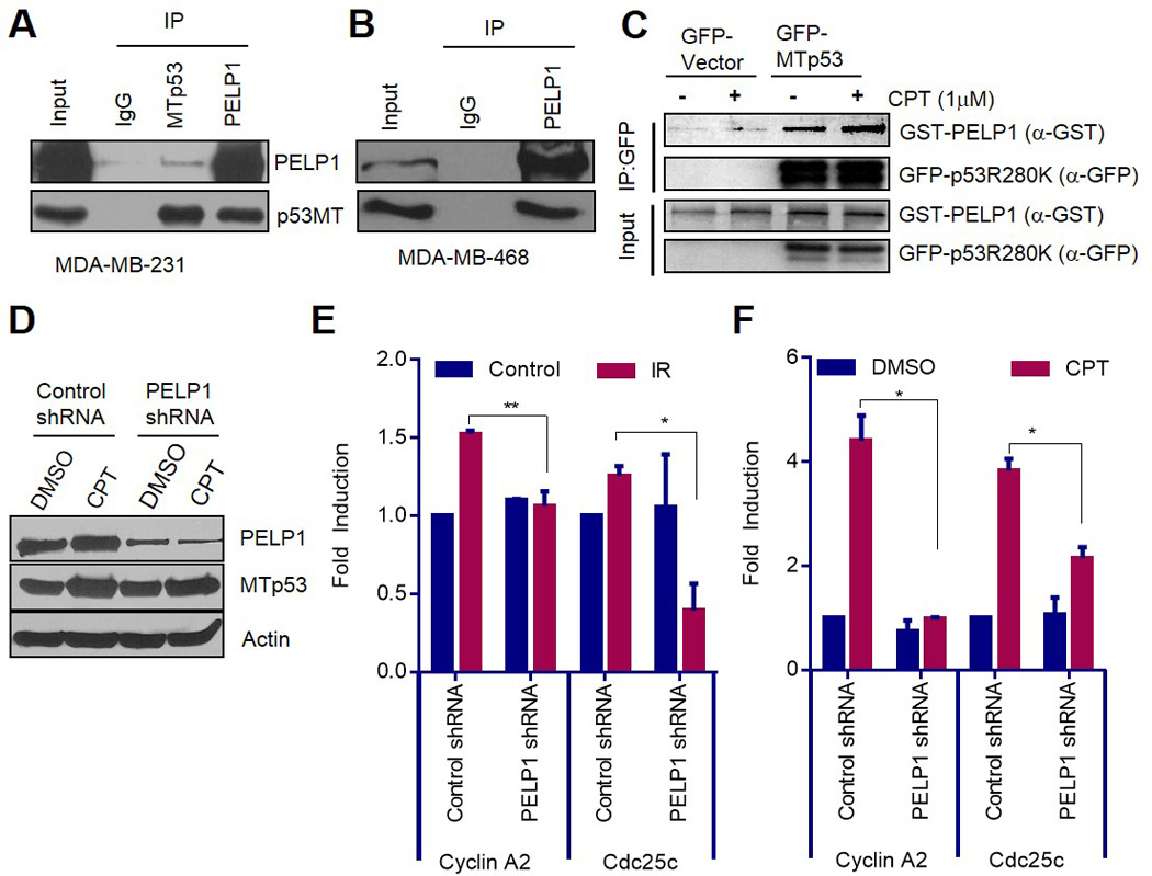
PELP1 interacts with MTp53 and regulates its target gene expression. (A, B) MDA-MB-231 cells (A) and MDA-MB-468 cells (B) were treated with 1µM of camptothecin for 6 h, and immunoprecipitation was done using IgG, PELP1, or p53 antibodies. Immunoprecipitates were subjected to Western blot analysis using PELP1 or p53 antibodies. (C) HEK-293T cells were transfected with GFP or GFP-MTp53 (R280K) along with GST-PELP1-expressing plasmids. After 48 h, cells were treated with 0.1% DMSO or 1µM of camptothecin for 2 h. GFP-trap IP and Western analysis was subsequently performed using GFP or GST antibodies. (D) MDA-MB-231 cells stably expressing control-shRNA or PELP1-shRNA were treated with 1 µM of camptothecin for 24 h and the expression of PELP1 and MTp53 was analyzed using Western analysis. (E, F) MDA-MB-231 cells were either exposed to 5gy gamma irradiation, and allowed to recover for 9 h (E) or treated with DMSO or 1µM of camptothecin for 24 h (F). cDNA synthesis and qRT-PCR analysis were subsequently performed to determine MTp53 target gene (Cdc25c and Cyclin A2) expression. *, P<0.05; **, P<0.01.