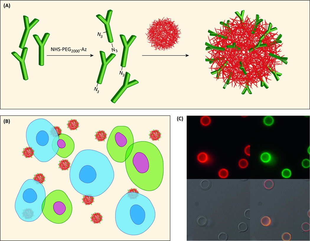
Figure 4.
Antibody functionalization and visualization. (a) Antibodies conjugated to the NP surface through “click” chemistry. (b) Cells that express the complementary antigen are blue and show Ab-facilitated binding of targeted NPs. Cells that do not express the complementary antigen are green with no NP binding. (c) Fluorescence microscopy images of huA33 mAbAzfunctionalized nanocapsules with (i) the antibody labeled with AF647 (red), or (ii) antibody labeled with AF488 (green), (iii) brightfield, and (iv) overlay images. Adapted with permission from [61].

