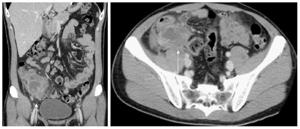
Figure 1.

Representative computed tomography images of appendiceal diverticulitis. An oblique coronal reformation of a contrast-enhanced CT (5-mm thick) (arrow) showing localized periappendiceal abscess. However, the appendix itself is not visualized clearly.
