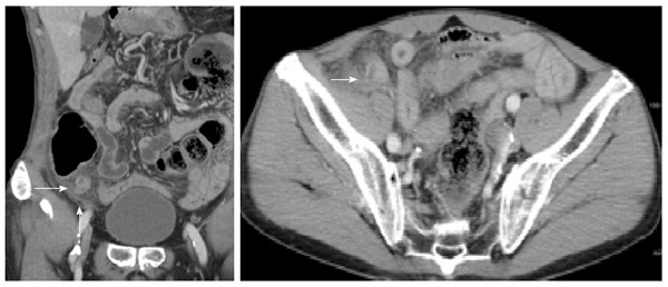
Figure 2.

Computed tomography scan showing appendiceal diverticulitis. An oblique coronal reformation of contrast-enhanced CT scan (5-mm thick) showing an inflamed diverticulum (arrow) and stranded surrounding fat. The inflamed diverticulum is visualized as a small, round cyst with an enhancing wall attached to the distal segment of the appendix. However, this patient’s appendix was not filled with fluid or enlarged.
