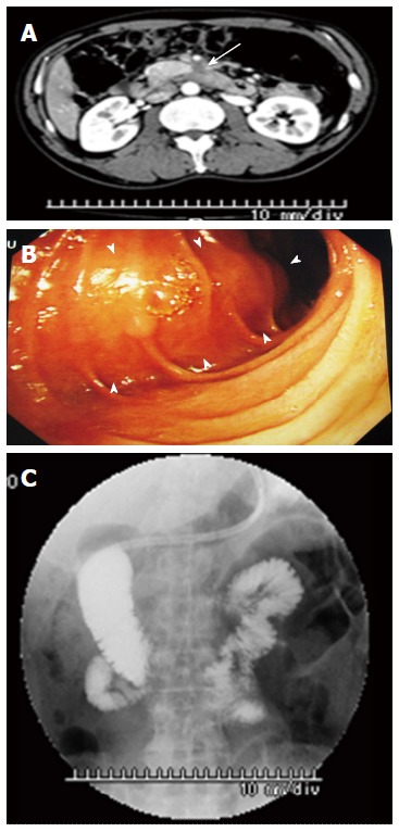
Figure 1.

Contrast-enhanced computed tomography, duodenofiberoscopy, and hypotonic duodenography. A: Abdominal CT scan shows a poorly enhanced mass around the third portion of the duodenum (arrow); B: Duodenofiberoscopic findings reveal submucosal tumors at the third portion of the duodenum (arrowheads); C: Hypotonic duodenography reveals stenosis at the third portion of the duodenum. CT: Computed tomography.
