
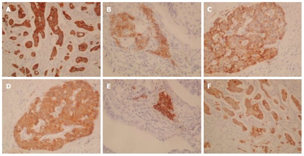
Figure 4.
Immunohistochemical staining: positive staining. A: Cytokeratin (CK) 7 (× 100); B: Neural cell adhesion molecule (CD56) (× 200); C: Chromogranin A (× 200); D: Synaptophysin (× 200); E: Insulin (× 200); F: Mucin 1 (MUC1).
Official websites use .gov
A
.gov website belongs to an official
government organization in the United States.
Secure .gov websites use HTTPS
A lock (
) or https:// means you've safely
connected to the .gov website. Share sensitive
information only on official, secure websites.
Immunohistochemical staining: positive staining. A: Cytokeratin (CK) 7 (× 100); B: Neural cell adhesion molecule (CD56) (× 200); C: Chromogranin A (× 200); D: Synaptophysin (× 200); E: Insulin (× 200); F: Mucin 1 (MUC1).