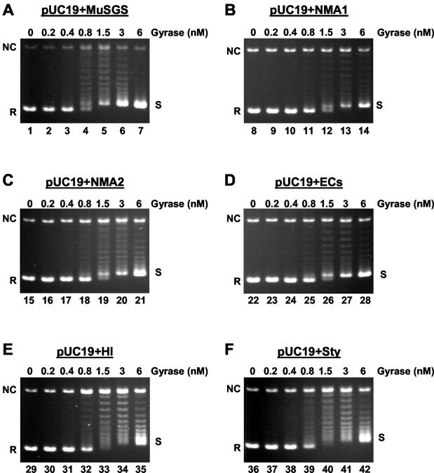
FIG. 4.
DNA gyrase-catalyzed supercoiling of 3-kb plasmids. Each 0.3-kb SGS homologue, along with the Mu SGS as a control, was cloned into pUC19 to generate six 3-kb substrates as indicated above each panel. Relaxed forms of these molecules (ca. 10 nM) were incubated as described in Materials and Methods, with increasing concentrations of DNA gyrase. The products were resolved on 1.1% agarose gels containing 45 μg of chloroquine/ml. On the gels presented, relaxed forms of the substrates run with the greatest mobility, indicated by R on the left of each panel. NC shows the position of nicked circular molecules, and the fully supercoiled topoisomers, shown as S on the right of each panel, run as an unresolved band closely behind the relaxed substrate.

