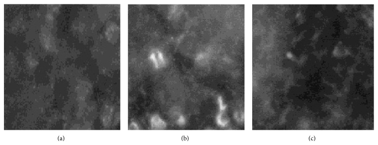
Figure 4.

Demonstration of iNOS protein in tumor. All photographs were taken at an exposure time of 1 s. Magnification ×400. (a) iNOS in cytoplasm of tumor cells of control rat. (b) iNOS in cytoplasm of tumor cells of IMO rat. (c) iNOS in cytoplasm of tumor cells of crocetin (40 mg/kg) rat.
