FIG 3.
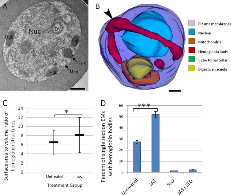
Effects of JAS treatment on hemoglobin trafficking in a trophozoite-stage IRBC. (A) Representative single-section electron micrograph of an IRBC treated with 7 μM JAS for 3 h. Note the appearance of multiple HCs (arrows) at the parasite periphery. Nuc, nucleus; Hgb, hemoglobin; Cyto, cytoplasm; Mito, mitochondria. (B) Representative 3D reconstruction of a JAS-treated IRBC. The hemoglobin-containing compartments, which appeared to be vesicles in a single-section electron micrograph, are actually part of a single large, hemoglobin-filled tube located at the periphery of the parasite. The tube lacks a cytostomal collar and is open to the erythrocyte cytosol (arrowhead). The parasite surface (dark blue), nucleus (light blue), DV (yellow), mitochondria (orange), hemoglobin-containing structures (red), and cytostomal collars (green) are shown. Scale bars, 500 nm. (C) Graph comparing the shapes of the HCs in untreated and JAS-treated IRBC. Untreated IRBC contain more spherical hemoglobin shapes, while the JAS-treated parasites have more oval HCs. (n = 13). (D) JAS treatment increases the percentage of IRBC with HCs approximately 2-fold. However, the number of HCv (as indicated by the percentage of single sections with hemoglobin after SLO treatment) remains the same with JAS treatment. n = 600. ***, P < 0.005; *, P < 0.05.

