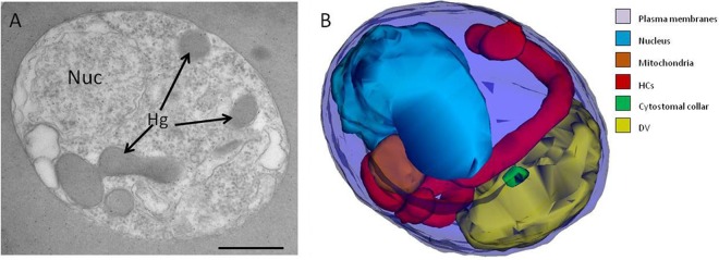
FIG 7.

NEM disrupts the hemoglobin transport pathway. (A) Representative single-section electron micrograph depicting a trophozoite-stage IRBC from a culture incubated with 4 mM NEM for 15 min. Nuc, nucleus; Hg, hemoglobin. (B) Typical 3D reconstruction of a NEM-treated trophozoite revealing the presence of a long, hemoglobin-filled tube without an electron-dense collar (red). Note the presence of a collar that is not associated with a cytostome (green). Scale bar, 500 nm.
