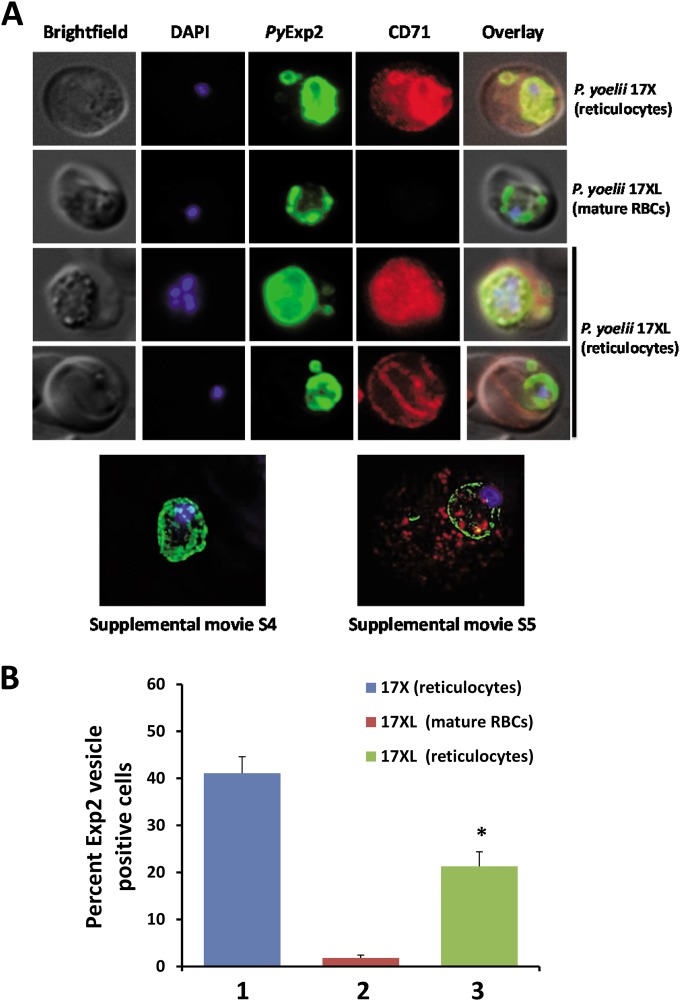
FIG 6.
PyExp2-positive vesicles are predominantly associated with reticulocytes infected with P. yoelii. (A) Immunofluorescence images showing costaining of PyExp2 (green) and CD71 (red) in RBCs infected with the P. yoelii 17X or P. yoelii 17XL strain. Shown at the bottom are representative sections of a 3D-SIM image of a P. yoelii 17XL-infected mature RBC (CD71 negative) and a P. yoelii 17XL-infected reticulocyte (CD71 positive); rotational 3D renderings are shown in Movies S4 and S5 in the supplemental material. (B) Percentages of infected cells containing PyExp2-positive vesicles in P. yoelii 17X-infected reticulocytes (1), P. yoelii 17XL-infected mature RBCs (2), or P. yoelii 17XL-infected reticulocytes (3). The percentages are expressed as means and SD. *, statistically significant difference (P < 0.05) between groups 2 and 3.

