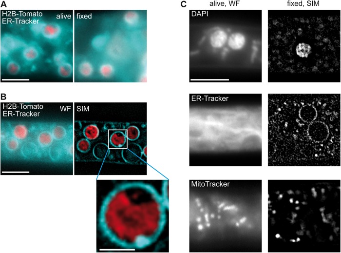
FIG 4.
Establishment of superresolution SIM for S. macrospora. (A) Hyphae of S. macrospora N883 carrying a tdTomato-tagged histone 2B (H2B) were stained with ER-Tracker. Unfixed cells (left) were compared to cells fixed with 0.2% formaldehyde (right). Bar, 5 μm. (B) To determine the effect of Fourier transformation, strain N883 was stained with ER-Tracker and micrographs were taken before (WF) and after (SIM) computational reconstruction. Bars, 5 μm and 1 μm (inset). (C) Dyes for different organelles were tested for use in SIM. Bar, 5 μm.

