
Figure 1. NA promotes autocrine production of TNFα and is required for necroptosis.
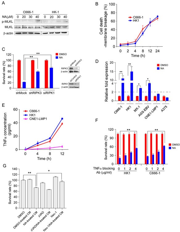
A. MLKL phosphorylation was detected using an MLKL phosphor-specific antibody. C666-1 and HK1 cells were treated with NA for 8 h and then harvested. Whole-cell lysates were subjected to SDS-PAGE followed by Western blot analysis. β-Actin is shown as a loading control. B. The number of dead cells was determined by measuring membrane integrity. C666-1 and HK1 cells were treated with NA and harvested at the indicated time points and membrane integrity was determined by trypan blue staining. C. RIPK1 and RIPK3 expression was knocked down in C666-1 cells, and then cells were treated with NA. Cell viability was estimated by MTS assay. D. NA treatment promotes TNFα transcription. Cells were treated with NA (40 μM) for 8 h and the TNFα mRNA level was determined by quantitative real time-PCR. E. NA triggers autocrine production of TNFα. Cells were treated as the indicated time points. Supernatant fractions from control and NA-stimulated cells were removed at the indicated time points and the secreted TNFα level was measured by ELISA. F. Autocrine signaling is required for NA-induced cell death. C666-1 cells were pre-treated (1 h) with neutralizing antibodies (1-4 μg/mL) against TNFα prior to treatment with 40 μM NA. Cell viability was estimated by MTS assay. G. Soluble factors mediate the anti-proliferation effect of NA. Cells were treated for 4 h with NA, washed with PBS 3 times, and fresh medium was added and cells incubated for another 6 h. At that time, the medium was collected as conditioned medium. Each graphical representation indicates the means ± S.D. of at least three independent testing conditions. *p<0.05. **p<0.001.