
Figure 4. The RIPK/NF-κB pathway mediates NA-induced TNFα production and cell death.cA.
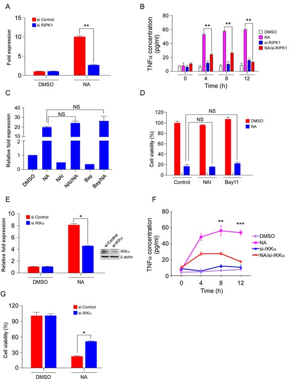
C666-1 cells transfected for 48 h with siRNA targeting RIPK1 or an empty vector control were treated or not treated with 40 μM NA for 8 h and relative levels of the TNFα transcript were determined and compared with β-actin and the fold change was calculated by comparing with DMSO-treated cells. B. C666-1 cells transfected for 48 h with siRNA targeting RIPK1 or an empty vector control were treated or not treated with 40 μM NA and harvested at the indicated time points. The presence of TNFα in conditioned cell culture media was measured by Elisa assay. C. The effect of NAI and Bay117082 (Bay) on NA-induced TNFα transcription. Cells were pre-treated with NAI (40 μM) and Bay117082 (Bay) (5 μM) for 1 h, and then treated or not treated with NA (40 μM). TNFα mRNA level was analyzed by quantitative-real time-PCR. D. Cells were pre-treated with NAI (40 μM) and Bay117082 (Bay) (5 μM) for 1 h, and then treated or not treated with NA (40 μM). Cell viability was estimated by MTS assay. E. IKKα in C666-1 cells was knocked down with siRNA, and then cells were treated with NA. TNFα mRNA level was analyzed by quantitative-real time-PCR. F. C666-1 cells transfected for 48 h with siRNA targeting IKKα or an empty vector control were treated or not treated with 40 μM NA and harvested at the indicated time points. The presence of TNFα in conditioned cell culture media was measured by Elisa assay. G. IKKα in C666-1 cells was knocked down with siRNA, and then cells were treated with different doses of NA. Viability of C666-1 cells was analyzed by MTS assay. Data are shown as means ± S.D. of values from three independent experiments. *p<0.05. **p<0.001. ***p<0.0001