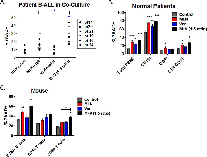
Figure 3. The combination of MLN0128/vorinostat increases killing of primary B-ALL cells with lesser effects on normal lymphocytes.

(A) Six non-Ph B-ALL patient specimens were cultured on stromal cells in the absence or presence of MLN0128 (100 nM), vorinostat (500 nM) or the combination. FACS was used to determine the percentage of hCD19+ cells that were non-viable (7-AAD-positive) after 48hr. (B) PBMCs from normal human donors were cultured for 48hr in media without cytokines. Cells were stained with anti-CD4 to mark helper T cells and anti-CD19 to mark B cells. Cytotoxicity was measured as the percent 7-AAD-positive as in Figure 3A. Viability of total PBMCs was determined using the ungated cells. Viability of CD4+, CD19+, and CD4-CD19- cells (which comprise mainly CD8+ T cells and natural killer cells) was determined using a lymphocyte gate based on forward and side scatter. Data represent mean +/− SEM (n = 5). (C) Purified mouse B-cells were cultured in BAFF and IL-4; purified T cells were cultured in IL-7 and IL-15. T cells were stained with anti-mouse CD4 and anti-mouse CD8 before viability analysis. Cytotoxicity was measured as the percent 7-AAD-positive as in panels A and B. For all panels, * p < 0.05; ** p < 0.01, unpaired two-tailed t-test.
