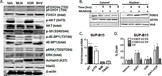
Figure 5. MLN0128 induces dephosphorylation and nuclear translocation of FOXO factors (A) Lysates from SUP-B15 cells treated for the indicated times with vehicle alone, MLN0128 (100 nM), vorinostat (500 nM), or the combination (M + V).

Blots were probed for the proteins and phosphosites shown on the right. (B) SUP-B15 cells were treated for the indicated times with MLN0128 (100 nM) before cell fractionation into nuclear and cytoplasmic extracts. Fractions were subjected to western blotting with anti-FOXO1 antibody. Antibodies to β-actin and histone H3 were used to confirm the purity of cytoplasmic and nuclear fractions. (C) SUP-B15 cells were treated for 4 hr with vehicle or 100 nM MLN0128. mRNA was obtained and expression of the indicated gene products was determined by Q-PCR. Graph depicts the fold increase in mRNA in cells treated with MLN0128 versus vehicle (mean +/− SEM, n = 3). *** p < 0.001, one-way ANOVA. BIM and TRAIL mRNA was not increased at 6 or 8 hr post-treatment (not shown). (D) SUP-B15 cells were infected with retroviruses expressing a human CD4 marker gene lacking the cytoplasmic tail and magnetically sorted to enrich hCD4+ cells. The vectors contained no insert (EV) or cDNAs encoding triple-alanine mutants of FOXO1 or FOXO3a fused to the hormone binding domain of the estrogen receptor (ER). Cells were then treated with vehicle or 4OHT for 48hr in the absence or presence of vorinostat (500 nM). The percentage of cell death was determined after 48 hr by 7-AAD staining (mean +/− SEM, n = 3). * p < 0.05; paired two-tailed t-test.
