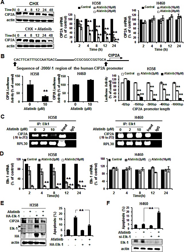
Figure 4. Elk-1 regulated CIP2A in NSCLC cells by afatinib.

(A) Left, H358 cells were treated with 100 μg/ml cycloheximide (CHX) in the presence or absence of afatinib for the indicated length of time. Middle and Right, H358 and H460 cells treated with afatinib at 10 μM or 20 μM for the designated incubation time. Afatinib inhibited CIP2A mRNA in a dose- and time-dependent manner, especially in H358 cells. Data are mean ± SD. n = 3 for each time point. *, p < 0.05, **, p < 0.01, vs. no afatinib. (B) Effects of afatinib on CIP2A promoter activity. Left, H358 and H460 cells were co-transfected with CIP2A reporter constructs (−1 to −2000bp) and renilla luciferase vectors for 48 h then treated with 10 μM afatinib for an additional 24 h. Afatinib decreased CIP2A luciferase activity in H358 cells, but not in H460 cells. Right, H358 cells were transfected CIP2A reporter various lengths of constructs and renilla luciferase vector for 48 h and then treated with 2 μM or 10 μM afatinib for an additional 24 h. Cell lysates were prepared for analysis of luciferase activity. Data are mean ± SD. n = 3 for each condition. **, p < 0.01, vs. no afatinib. (C) Chromatin immunoprecipitation assays of the CIP2A promoter. H358 and H460 cells were treated with 2 μM or 10 μM afatinib for 24 h and processed for ChIP assay. Soluble chromatin was immunoprecipitated with specific Elk-1 or IgG (negative control) antibodies. Immunoprecipitates were subjected to PCR with primer pair specific to CIP2A promoter (−16 to −213 bp) and RPL30 (internal control). The gel shown is representative of three independent experiments. (D) H358 and H460 cells treated with afatinib at 2 μM or 10 μM for the indicated incubation times. Afatinib inhibited Elk-1 mRNA in a dose- and time-dependent manner, especially in H358 cells. Data are mean ± SD. n = 3 for each time point. **, p < 0.01, vs. no afatinib. (E) Ectopic expression of Elk1 (HA-Elk1) restored the effect of afatinib on CIP2A expression and protected the effect of afatinib-induced apoptosis in H358 cells using Western blotting and FACS. H358 cells overexpressing Elk-1 were treated with 10 μM afatinib for 24 h. Open arrow is endogenous of Elk1 and close arrow is exogenous of Elk1. (F) Knockdown of Elk-1 enhanced apoptosis in H460 cells by afatinib. Protein levels of Elk1 expressed in lower panel. Data are means ± SD. **, p < 0.01.
