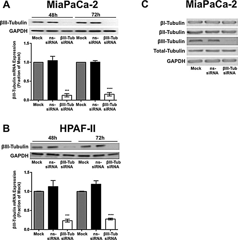
Figure 2. βIII-tubulin silencing in pancreatic cancer cell lines.
A) Top panel, Western blot analysis of βIII-tubulin silencing in protein extracts from MiaPaCa-2 cells. Cell lysates were harvested from cells 48h or 72h after transfection with mock, control siRNA (ns-siRNA), or βIII-tubulin siRNA (βIII-Tub siRNA). GAPDH was used as a loading control. Bottom graph, real-time PCR analysis of βIII-tubulin silencing in MiaPaCa-2 cells. RNA was harvested from cells 48h or 72h after transfection with mock, ns-siRNA, or βIII-tub siRNA. βIII-tubulin mRNA levels were normalized to 18S mRNA. B) as per A, except cell extracts were obtained from HPAF-II cells. Asterisks indicate significance (** p≤0.01, ** p≤0.01; n=3). C) Representative Western blots for βI-, βII-, βIII-tubulin and total tubulin in protein extracts from MiaPaCa-2 cells transfected with mock, ns-siRNA, or βIII-Tub siRNA (n=3). GAPDH was used as a loading control.

