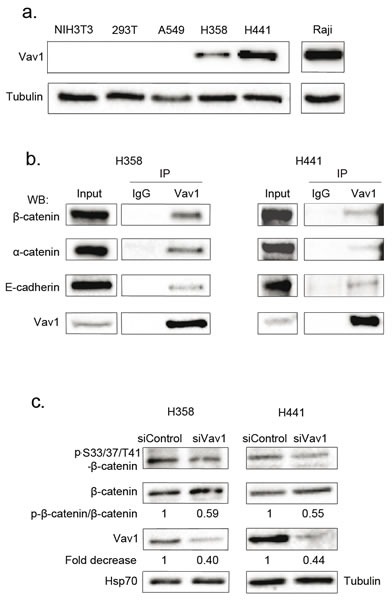
Figure 7. Vav1 interacts with β-catenin in lung epithelial cancer cell lines.

a. Vav1 protein expression in pulmonary and lymphoid cell lines. Total cell extracts were analysed by sequential immunoblotting with the indicated Abs. b. Total cell extracts from the indicated cells were immunoprecipitated (IP) with anti-Vav1 Ab or control IgG. Immune complexes or total extracts (Input) were analysed by sequential immunoblotting with the indicated Abs. c. Η358 and Η441 cells were transfected with the indicated siRNA and proteins levels assessed 72 hours after transfection. Ratio between phospho-β-catenin and total β-catenin was calculated relative to transfection of control siRNA.
