Fig. 6.
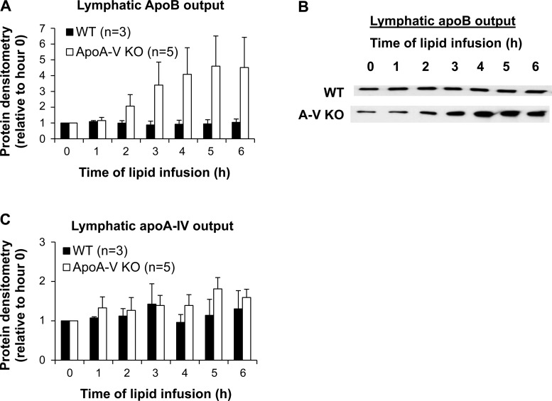
ApoA-V KO mice have increased intestinal apoB secretion into the lymph. A: lymph samples were analyzed for apoB by Western blot. Protein concentration was quantified by densitometric analysis and normalized to hour 0 (fasting). B: a representative blot is shown for lymphatic apoB output. C: lymph samples were analyzed for apoA-IV by Western blot.

